Nudibranches are often studied by neurobiologists because they have very large, identifiable, neurons that are very amenable to neurophysiological investigations. Our studies of the nudibranch, Melibe leonina, center around their rhythmic behaviors, such as feeding and swimming. We seek to identify the neural circuits that generate the behaviors as well as the neurons and hormones that modulate the expression of these behaviors.

Melibe Swimming
A number of different molluscs swim and the neural basis of swimming has been investigated in several different species, such as Clione, Tritonia, Pleurobranchaea and Aplysia brasiliana. Swimming in Melibe is bit different from these species because they use lateral bending motions to propel themselves (see figure below and video), rather than flapping their parapodia or undulating in a dorsal-ventral direction.
To see video of a Melibe swimming click on the link below:
Melibe swim spontaneously, and in response to contact with a potential predator, such as a starfish. Spontaneous swimming appears to be influenced by light, and time of day.
Further details about Melibe swimming behavior can be found in the following publications:
W.H. Watson III, K.D. Lawrence and J.M. Newcomb. 2001. Neuroethology of Melibe leonina swimming behavior. American Zoologist 41(4): 1026-1035. doi.org/10.1093/icb/41.4.1026
Newcomb, J.M. and W.H. Watson III. 2001. Modification of swimming in the gastropod Melibe leonine by nitric oxide. J. exp. Biol. 205: 397-403.
Lawrence, K.A. and W.H. Watson III. 2002. Swimming behavior of the nudibranch Melibe leonine. Biol. Bull. 203: 144-151.
Newcomb, J.M., K.D. Lawrence and W.H. Watson III. 2005. The influence of light on locomotion in the gastropod Melibe leonina . Marine and Freshwater Behav. & Physiol. 37: 253-269. https://doi.org/10.1080/10236240400016629

Swimming, in intact animals and isolated brains, appears to be inhibited by light.

We, and others, most notably members of Paul Katz's lab, have studied the neural basis of Melibe swimming behavior in detail. Using both semi-intact and isolated brain preparations we have been able to identify the neurons involved and determine how they generate the swimming rhythm.
In order to produce a rhythmic behavior, such as swimming, feeding and walking, neurons are wired together into central pattern generating networks or CPGs. Invertebrate nervous systems are particularly well suited for studies of CPGs because discrete ganglia can be studied in vitro and the neurons in these ganglia are very large and identifiable. This makes it possible to investigate the same cells and networks from one animal to the next.
We have now identified the CPGs in the brain and buccal ganglia which produce the swimming and swallowing behaviors but the location of the feeding CPG is still in question. Some of the neurons that produce swimming are shown in the figures below. Once we identified the neural networks that produce these behaviors, it was possible to study how these networks interact with each other (behavioral choice) and how they are modulated and turned on and off by neuropeptides such as SCP.
A brief summary is provided below and additional details can be found in the following papers:
Watson, W.H. III, J.A. Newcomb and S. Thompson. 2002. Neural correlates of swimming behavior in Melibe leonina. Biol. Bull. 203: 152-160.
Thompson, S. and W. H. Watson III. 2005. Central pattern generator for swimming in Melibe J. Exp. Biol. 208:1347-1361. https://doi.org/10.1242/jeb.01500
Semi-Intact Preparations
It is possible to obtain intracellular recordings from "freely behaving" Melibe. Under these circumstances cells in each pedal ganglia fire in phase with the lateral body movements that are characteristic of swimming. Firing of swim motoneurons in the left pedal ganglia causes the animal to bend to the left and visa versa.

Isolated brain
The isolated Melibe brainwill spontaneously produce the swimming motor program.

Light inhibits expression of the pattern.
Swim CPG
The swim central pattern generator (CPG) consists, at a minimum, of a pair of cells in the cerebropleural ganglion (left and right Interneuron I) and a pair of cells in the pedal ganglia (left and right Interneuron II). The anatomy of these cells is shown below.
The two cells on one side of the brain inhibit the pair on the other side, and the cells on a given side are electrically coupled to each other. See below for schematic of swim circuit.

Inhibition of any cell in the CPG stops the swim rhythm and stimulation of any cell in a nonswimming preparation or semi-intact animal initiates swimming.
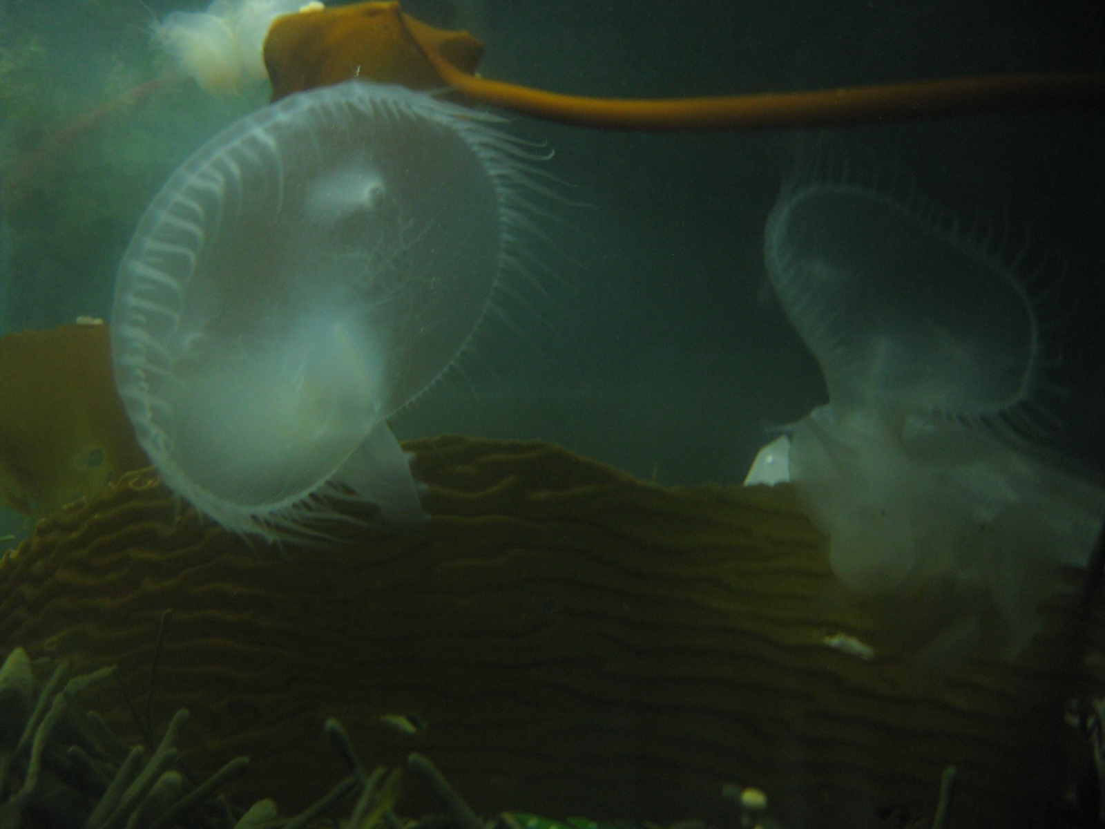
Melibe feed with rhythmic movements of their oral hood (see video below). In the image below both Melibe have their oral hoods fully open. When they close their oral hoods they will engulf a bolus of seawater and they will contracts the oral hood to push water out their a type of sieve made with their tentacles. This allows them to capture small zooplankton. Sometimes they also use the same motion to kind of scrap algae and small organisms off of kelp or seagrass blades.
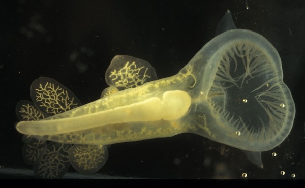
Unlike many nudibranchs, Melibe does not chew its food. Rather it simply swallows things it engulf using parastalis of the esophagus, controlled by their buccal ganglia. Food then enters both their stomach and digestive diverticuli, which are very branches and run along the inside of their translucent skin. This enables the Melibe to take on the color of their environment and perhaps blend in better. An image of a Melibe below shows their diverticuli in more detail.
The following papers provide further details about Melibe feeding behavior:
Watson, W.H. and J.Trimarchi. 1992. A quantitative description of Melibe feeding behavior and its modification by prey density. Marine Behaviour and Physiology 19: 183-194.
Trimarchi, J. and W.H. Watson. 1992. The role of the Melibe buccal ganglia in feeding behavior. Marine Behaviour and Physiology 19: 195-209.
Watson, W.H. III. and C.M. Chester. 1993. The influence of olfactory and tactile stimuli on the feeding behavior of Melibe leonina (Gould, 1852) (Opisthobranchia: Dendronotacea). The Veliger 36(4): 311-316.
Watson, W.H. III, K. M. F. Bourque,J. R. Sullivan, M. Miller, A. Buell, M. G. Kallins, E. Curtis, S. K. Pierce, E. Blackman, S. Urato and J. M. Newcomb. 2017. The digestive diverticula in the carnivorous nudibranch, Melibe leonina, do not contain photosynthetic symbionts. Integ. Org. Biol. 3(1): 1-11. doi:10.1093/iob/obab015
Melibe feed using rhythmic movements of their oral hood. However, the neural network responsible for generating the rhythm has not been elucidated.
After feeding for about 30 minutes Melibe typically become satiated and stop. This is due to feedback from the stomach to both the brain and the buccal ganglia. Inflation of the stomach led to satiation and cutting the nerves that carry information from the stomach to the buccal ganglia prevented satiation.
For more information see:
Lee, C and WH Watson III. 2016. The influence of stomach distension on feeding in the nudibranch mollusk Melibe leonina. Marine and Freshwater Behav. and Physiol. 49 (4): 277-290. doi.org/10.1080/10236244.2016.1192305.
Nitric oxide is a gaseous neurotransmitter or neuromodulator produced by the enzyme nitric oxide synthase, in response to calcium. In Melibe NOS is found in two neurons in the brain and these cells project to the pedal ganglia, where many neurons controlling locomotion and swimming are located. Treatment of isolated brains, which spontaneously produce a swimming rhythm, with NO donors, slows and inhibits the rhythm. Stimulation of the NOS neurons has similar effects. These data are summarized, with figures, below.
Anatomy I. Diaphorase staining
Histochemical staining of the Melibe brain using the NADPH-diaphorase method reveals only two bilaterally symmetricl neurons in the cerebropleural ganglion.

These two cells do not stain if the appropriate controls are performed, or if NOS is blocked.
Interestingly, Melibe tentacles also stain for NOS, although the significance of the observation is not clear at the present time.
Anatomy II. Immunohistochemistry
Immunohistochemical staining of Melibe brains with anti-NOS antibodies results in staining of the same two neurons that are revealed with diaphorase histochemistry.

Anatomy III. Lucifer yellow injections
Injection of the NOS neurons with lucifer yellow revealed more detail about their structure. Double labelling with diaphorase confirmed that we injected the correct cells.

Pharmacology and Physiology of NO in Melibe
Treatment of isolated Melibe brains with a NO-donor, such as sodium nitroprusside (SNP) alters the normal swim rhythm. It slows considerably and becomes more erratic.

In intact animals this manifests itself as very slow swimming. The effect of NO donors is eliminated by perfusion with the NO scavenger hemoglobin. Theses same effects were obtained with cGMP analogues.
Stimulation of the NOS neuron also inhibits the swim rhythm.

Melibe move by either crawling or swimming and they tend to do both more during the night. The following paper provides further details about Melibe locomotion and their tendency to be nocturnal.
Newcomb, J.M., L.E. Kirouac, A.A. Naimie, K.A. Bixby, C. Lee, S. Malanga, M. Raubach and W.H. Watson III. 2014. Circadian rhythms of crawling and swimming in the nudibranch mollusk Melibe leonina. Biol. Bull. 227: 263-273. doi.org/10.1086/BBLv227n3p26